


What to Expect at Your 7-Week Ultrasound Appointment

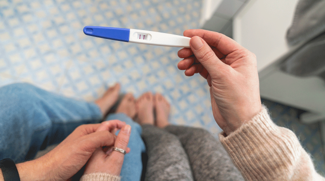
There’s nothing quite as satisfying as seeing your fast-growing baby—and the flicker of their little heartbeat—on an ultrasound scan. If you just got your positive pregnancy test (Congrats!), you might be wondering if you need an ultrasound around 7 weeks.
The American College of Obstetricians and Gynecologists (ACOG) officially recommends at least one ultrasound during pregnancy—the mid-pregnancy ultrasound at 18 to 22 weeks—unless you’re at risk for certain complications. But in practice, many providers offer more than one ultrasound—including one in the first trimester.
“I had an ultrasound at 7 weeks because I had just found out I was pregnant and made my first prenatal appointment as fast as possible since I was so excited,” shares Natalie Gontcharova, senior editor at The Bump and mom of one.
Here, learn why your provider might order a 7-week ultrasound and what to expect.
It’s relatively uncommon to get an ultrasound at 7 weeks, says Catherine Caponero, DO, an ob-gyn at Cleveland Clinic. Some practitioners, however, do perform a first-trimester ultrasound starting at 7 weeks of pregnancy, since that’s when a heartbeat should be visible, to confirm viability and estimate your due date—particularly if you aren’t sure of the date of your last menstrual period or have irregular periods, or if you had complications in a previous pregnancy.
First trimester scans can also be helpful in determining genetic abnormalities. However, most of the time these scans are performed later. “Most of our first trimester scans are done between 11 to 13.6 weeks, for the nuchal translucency,” says Kimberly Khabeer, a sonographer at Temple University Hospital in Philadelphia.
You might get a 7-week ultrasound to confirm the embryo is located inside the uterus and has a heartbeat. Your doctor may also want to monitor for potential problems, including to check for an ectopic pregnancy (when the embryo implants outside of the uterus), evaluate vaginal bleeding, figure out the cause for pelvic pain, diagnose multiple gestations or locate or remove an intrauterine device (IUD), says Caponero.
I went in at what I thought was 8 weeks, 2 days and got an ultrasound and based on the results, they dated me at 7 weeks 3 days. I could see the sac/placenta beginning and the adorable little peanut with a heartbeat. It looks like a little tiny flutter just going crazy. It was the best thing I've ever seen. I got to bring home the first ‘pictures’ of my little peanut. I didn't think we’d see anything, but (usually) you can and it was thrilling.
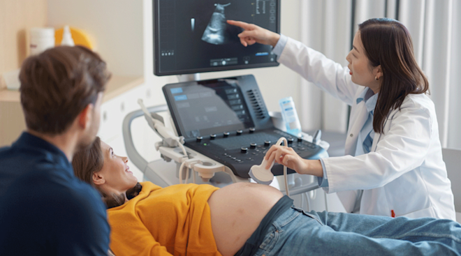
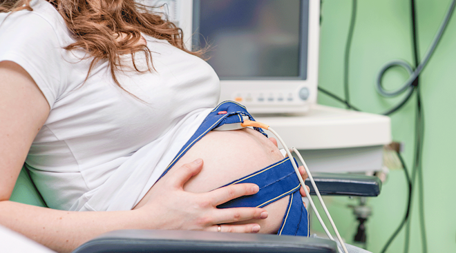
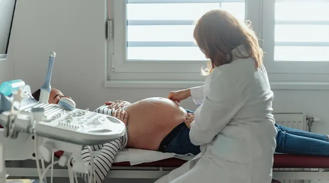
During a 7-week ultrasound, your practitioner may attempt a transabdominal ultrasound, where the wand is placed on your lower abdomen. If they can’t see what they need to see, they’ll give you a transvaginal ultrasound, where the wand is placed inside the vagina.
Your healthcare provider will examine your uterus, cervix and the area around the ovaries and fallopian tubes, to make sure everything looks as expected. They’ll check for the location of a gestational sac (a fluid-filled structure surrounding the embryo) and yolk sac (which nourishes the pregnancy until the placenta takes over at around 12 weeks). They’ll also measure the crown-to-rump (head-to-butt) length of the embryo and check cardiac activity.
You don’t necessarily need to prepare for this appointment. But if your doctor wants to attempt a transabdominal ultrasound, they may ask that you come to your appointment with a full bladder, which makes it easier to see things.
I had my high-risk consultation at 7 weeks, 1 day and they did an ultrasound to date the pregnancy, even though I knew the dates perfectly. We saw the little baby and the heartbeat! Mine was transvaginal, in part because I’m fat and because it was so early anyway. Abdominally we did see the gestational sac.
At 7 weeks pregnant, baby’s the size of a blueberry. An ultrasound will usually show a gestational sac, yolk sac and a small embryo. “It might resemble a grain of rice with flickering of the cardiac cords,” says Khabeer.
Can twins be detected at a 7-week ultrasound?
While twins can be detected at a 7-week ultrasound, they can be missed depending on where the pregnancy is located in the uterus, the type of twins or even your anatomy, says Caponero.
What if you see nothing at the 7-week ultrasound?
It’s possible that you won’t see a gestational sac or an embryo at your 7-week ultrasound even though you’re pregnant. That’s because it can be challenging to accurately date a pregnancy early on.
That said, at 7 weeks, your provider can diagnose a miscarriage “if the gestational sac is too big and an embryo isn’t seen, or if the embryo doesn’t have a heartbeat and is at least 7 millimeters long,” says Caponero. They also may suspect an ectopic pregnancy if they don’t see a gestational sac inside of the uterus. If that happens, your practitioner will check around the uterus for any abnormalities to figure out the best next steps.
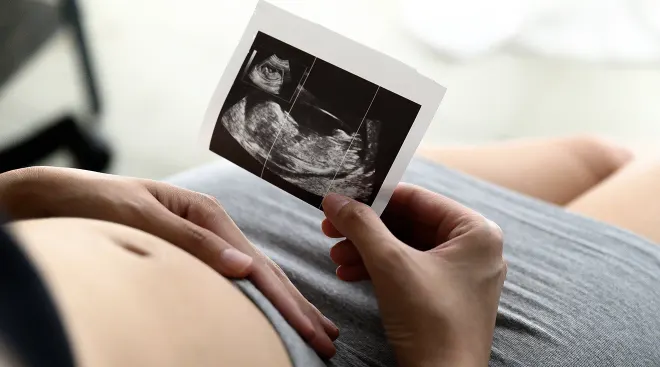
If you get an ultrasound at 7 weeks, your provider or technician will let you get a sneak peek of the screen and print out a blurry photo for you. You might not be able to tell what’s what, but it might look something like this:
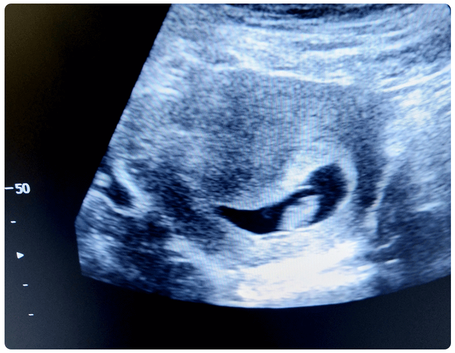
My 7-week ultrasound looked like a little white blob with an even smaller blob next to it.
After a 7-week ultrasound, next steps depend on what your practitioner observes. If they only see a gestational sac inside the uterus, the dating of your pregnancy may have been off. Your doctor will often recommend waiting two weeks before repeating an ultrasound, at which point they should be able to see an embryo with a heartbeat. “While it can be very hard to wait, it’s so important for expectant parents to have a clear answer instead of uncertain possibilities,” says Caponero. If your practitioner doesn’t see a gestational sac in your uterus, they may order bloodwork to determine the best timing for additional imaging, testing or treatment, Caponero says.
Frequently Asked Questions
What are the odds of miscarriage after seeing a heartbeat at 7 weeks?
Seeing a heartbeat significantly reduces the odds of miscarriage, especially as your pregnancy progresses. If your practitioner sees a heartbeat at your 7-week scan, your odds of miscarriage are relatively low—only about 4.2 percent, according to one study.
Can you have a 3D ultrasound at 7 weeks pregnant?
It’s possible to have a 3D ultrasound, but your provider may not recommend it. “Unless there’s a medical need, most providers may not recommend a 3D ultrasound,” says Caponero, to avoid unnecessary interventions.
Do midwives perform regular ultrasounds?
Yes, some midwives can and do perform ultrasounds. “Our midwife team does a wonderful job evaluating patients and completing first trimester ultrasounds,” says Caponero.
How does your stomach look and feel at 7 weeks?
Remember, baby’s the size of a blueberry at 7 weeks pregnant, which means the uterus isn’t significantly bigger yet. Your stomach might feel a bit bloated and look slightly swollen, due to higher levels of the hormone progesterone.
“Cramping is also common at this time,” says Caponero. Call your doctor right away if you experience severe pain or heavy bleeding, or if you feel lightheaded, dizzy or weak. “This may be an indication that the pregnancy’s not in the correct location,” she adds, in which case “an ultrasound is an incredibly helpful diagnostic tool.”
What’s happening with baby’s development at 7 weeks?
At 7 weeks pregnant, baby’s brain is growing fast—gaining about 100 new cells every minute—and the spinal cord is forming (a big reason to take your prenatal vitamin!). Baby’s arm and leg buds now have some cartilage, and the lungs and digestive tract are continuing to develop. Although it won’t be obvious on your ultrasound, baby’s eyes, nostrils, ears and mouth are becoming more visible. Pretty soon, you’ll see hints of a baby on your ultrasound, rather than just a blob!
While it’s not super common, some practitioners perform an ultrasound starting at around 7 weeks to date the pregnancy and check for a heartbeat. Otherwise, a 7-week ultrasound is usually only recommended if your doctor wants to check out potential complications like pain and bleeding.
Please note: The Bump and the materials and information it contains are not intended to, and do not constitute, medical or other health advice or diagnosis and should not be used as such. You should always consult with a qualified physician or health professional about your specific circumstances.
Plus, more from The Bump:
Catherine Caponero, DO, is an ob-gyn at Cleveland Clinic. She earned her medical degree from Lake Erie College of Osteopathic Medicine in Erie, Pennsylvania.
Kimberly Khabeer is a sonographer at Temple University Hospital in Philadelphia.
American College of Obstetricians and Gynecologists, Ultrasound Exams, January 2024
Cleveland Clinic, Transvaginal Ultrasound, May 2022
Cleveland Clinic, Ultrasound in Pregnancy, September 2022
Cleveland Clinic, Yolk Sac, February 2022
Medline Plus, National Library of Medicine, Ultrasound Pregnancy, March 2024
Miscarriage Association, Ultrasound Scans
National Health Service UK, You and Your Baby at 7 Weeks Pregnant, October 2021
Nemours KidsHealth, Pregnancy Calendar: Week 7
Obstetrics & Gynecology, Miscarriage Risk for Asymptomatic Women After a Normal First-Trimester Prenatal Visit, March 2008
StatPearls, National Library of Medicine, Gestational Sac Evaluation, July 2023
StatPearls, National Library of Medicine, Sonography 1st Trimester Assessment, Protocols, and Interpretation, July 2023
Real-parent perspectives
- Natalie Gontcharova, senior editor at The Bump and mom of one
- lizparkerdeken, The Bump community member
- punkrockabye, The Bump community member
Learn how we ensure the accuracy of our content through our editorial and medical review process
Navigate forward to interact with the calendar and select a date. Press the question mark key to get the keyboard shortcuts for changing dates.