


What to Expect From the 6-Week Ultrasound

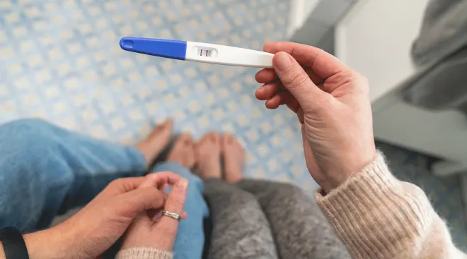
Just got a positive result on an at-home pregnancy test? You might be wondering if it’s time to schedule an ultrasound. But exactly when your doctor will recommend a first ultrasound depends on a few different factors. While they sometimes schedule a 6 week ultrasound for low-risk patients, they often suggest waiting until you’re at least 8 weeks along. Ahead, learn why your healthcare provider might recommend an ultrasound at 6 weeks—and what else to know about this prenatal test.
The American College of Obstetricians and Gynecologists (ACOG) recommends at least one scan during pregnancy, typically the mid-pregnancy ultrasound performed between 18 and 22 weeks. But, in practice, most providers offer three or four ultrasounds for their low-risk pregnant patients, including one between 6 to 10 weeks to confirm fetal viability, explains Mahino Talib, MD, an ob-gyn at NYU Langone Health and clinical assistant professor at NYU Grossman School of Medicine in New York City.
Maris Toland, MD, an ob-gyn at Dartmouth Hitchcock Clinics Specialty Care in Bedford, New Hampshire, says the number of ultrasounds you can expect depends on local imaging resources and your specific medical concerns. “For instance, if someone has vaginal bleeding or pain in early pregnancy, an ultrasound may be ordered to investigate for causes,” she says.
Between 6 to 10 weeks of pregnancy, practitioners often perform a “dating ultrasound” to confirm your estimated due date, confirm viability and make sure the pregnancy is “contained within the uterus,” says Talib. Doctors may also recommend a 6 week ultrasound if you’re experiencing bleeding or have had a previous ectopic pregnancy or C-section. These factors increase the risk of ectopic pregnancy, when the embryo implants outside of the uterus, which can be life-threatening, says Toland—so it’s important to diagnose and treat as soon as possible.
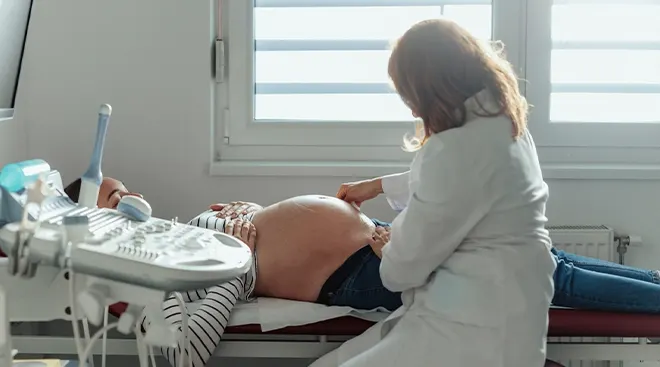
To ensure accuracy, a 6 week ultrasound is always performed transvaginally. This means that an ultrasound wand (known as a transducer) will be gently inserted into your vagina, instead of on top of your abdomen. “Six-week embryos are only about the size of a lentil,” says Toland. “They’re too small to be seen clearly with an ultrasound probe on the abdomen.”
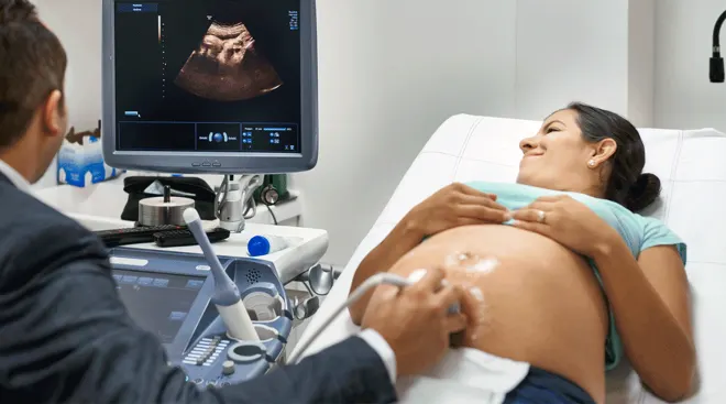
A transducer makes sound waves that bounce off structures inside the uterus to create images of the embryo that are projected onto a screen. It produces a sharper image when the transducer is inside the vagina, closer to your uterus. “Once a pregnancy is about 10 to 12 weeks, about the size of a strawberry, it can be seen well enough with a probe on the abdomen,” says Toland.
To perform a transvaginal ultrasound, your practitioner will wrap the transducer in a condom and apply a gel before inserting the wand in your vagina and gently moving it to view various structures. It may feel a bit uncomfortable, but it shouldn’t feel painful. The entire scan should take about 15 minutes to an hour.
Unless you have a medically trained eye, you won’t be able to identify much. To patients, the pregnancy will probably resemble a peanut at a 6 week ultrasound, says Talib.
Your ultrasound technician or practitioner’s trained eye will be able to identify a gestational sac (a fluid-filled pouch that’ll eventually become the amniotic sac), a yolk sac (a temporary structure that helps the pregnancy grow) and an embryo. “This is before any recognizable organs or limbs have developed,” says Toland. Your doctor will also check for ovarian cysts and uterine fibroids, Talib adds.
Will a heartbeat be detectable at the 6 week ultrasound?
Your practitioner may or may not be able to detect a flicker suggesting the electrical activity of the primitive fetal heart at the 6 week ultrasound. This is often referred to as a heartbeat, although the organ doesn’t beat in the way we typically imagine. “There are rhythm cells that are developing, so we do see a flicker on ultrasound that is typically referred to as cardiac activity or cardiac motion,” says Toland.
It’s also possible that you won’t see a heartbeat because your estimated due date is off. It’s usually not possible to know exactly when you ovulated and egg met sperm, so your estimated due date is initially based off of your last menstrual period. That means you could actually have booked your ultrasound at 5 weeks pregnant—at which point you wouldn’t see a heartbeat. “Not all patients have regular or reliable menstrual periods,” says Toland. “This is one reason why early first-trimester ultrasounds can be so helpful in accurately estimating the due date of a pregnancy.”
You also may not see a heartbeat if the pregnancy is ectopic (located outside of the uterus), or if the pregnancy “is headed in the direction of a miscarriage,” says Talib. Miscarriage affects about 10 to 20 percent of pregnancies, according to Mayo Clinic, and usually happens because of a chromosomal abnormality.
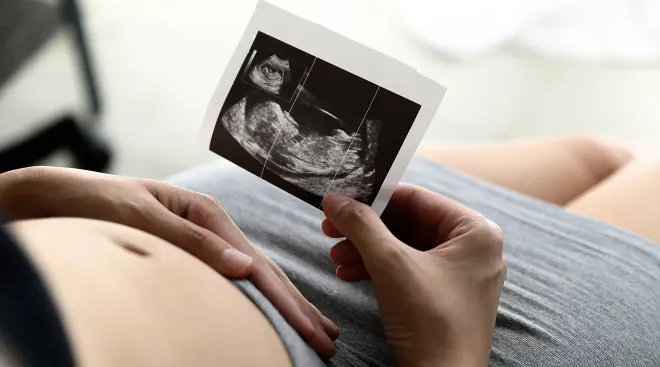
6 week ultrasound pictures
This is what a typical 6 week ultrasound looks like:

A 6 week ultrasound poses no health risks for you or baby. That said, technicians limit the duration of scans to reduce any theoretical risk to the embryo, says Toland—although there’s no evidence of fetal harm linked to diagnostic ultrasounds. Another risk: If you don’t see a heartbeat because you’re actually not yet 6 weeks pregnant, “it can cause undue stress,” adds Talib.
Some people find transvaginal ultrasounds physically or emotionally uncomfortable, especially if you have a history of trauma. If you’re concerned, Toland suggests bringing a supportive friend, partner or family member to your appointment. You may also prefer to place the transducer yourself. “Many ultrasound techs offer this. If they don’t, you can always ask upfront,” she says.
Before scheduling your 6 week ultrasound, try to accurately estimate your due date and check in with your doctor, to be sure you don’t waste your resources on a scan you don’t need. If your provider orders a transabdominal ultrasound, make sure to come with a full bladder—it’ll be much easier for the technician to see the images, says Toland.
Frequently Asked Questions
Does everyone get an ultrasound at 6 weeks?
Not everyone gets a 6 week ultrasound; it depends on where you live and your personal risk factors. Some doctors only perform one ultrasound, at around 18 to 22 weeks of pregnancy, unless you have risk factors (such as bleeding). That said, many practitioners do regularly schedule an early ultrasound between 6 to 10 weeks of pregnancy, to confirm the due date and location of the pregnancy.
Is 6 weeks early for an ultrasound?
To avoid stress for the patient, many practices recommend waiting until 8 weeks for a first ultrasound, unless you have pain, bleeding or other concerns, says Talib. (Ultrasounds typically aren’t performed before 6 weeks, since it isn’t possible to detect cardiac activity earlier in a pregnancy.)
If no heartbeat was detected at 6 weeks, will I have a follow up ultrasound?
If your practitioner doesn’t detect a heartbeat, they’ll usually repeat an ultrasound within 10 to 14 days. “At that point, if the pregnancy is viable, we should see a fetus with a heartbeat,” says Talib.
Will I also get an 8-week ultrasound?
You’ll likely only get an 8-week ultrasound if a 6-week ultrasound appears abnormal or if there are any lingering concerns—i.e., if there’s “no heartbeat yet, or the heartbeat appears slow,” says Talib.
When will my next ultrasound be?
Your doctor may offer a nuchal translucency ultrasound in roughly one month, between 11 to 14 weeks of pregnancy, to screen for chromosomal or cardiac abnormalities, says Talib.
Otherwise, in an uncomplicated pregnancy your next ultrasound will happen at around 18 to 22 weeks. Your provider will check that baby’s organs are developing as expected—and you’ll find out baby’s sex if you haven’t already. Some practitioners also offer a “sneak peek” ultrasound at around 36 weeks, says Talib, to double-check baby’s due date, verify their position and estimate their weight. You may get additional ultrasounds in the second and third trimesters if you have complications, “particularly if there are any concerns about the fetus’ size,” says Toland.
What’s happening with baby’s development at 6 weeks?
At 6 weeks of pregnancy, baby’s roughly the size of a sweet pea. They’re beginning to develop a brain and liver, as well as musculoskeletal, digestive and respiratory systems. They’re also growing arm and leg buds, as well as optic vessels and the inner ear.
While 6 week ultrasounds aren’t necessarily common, doctors do perform scans this early for low-risk pregnancies. That said, unless you have the above-mentioned risks, you may want to wait until the eight-week mark for your first ultrasound to avoid unnecessary stress.
Please note: The Bump and the materials and information it contains are not intended to, and do not constitute, medical or other health advice or diagnosis and should not be used as such. You should always consult with a qualified physician or health professional about your specific circumstances.
Plus, more from The Bump:
Mahino Talib, MD, is an ob-gyn at NYU Langone Health and clinical assistant professor at NYU Grossman School of Medicine in New York City. She earned her medical degree from American University of the Caribbean School of Medicine.
Maris K. Toland, MD, is an ob-gyn at Dartmouth Hitchcock Clinics Specialty Care in Bedford, New Hampshire. She earned her medical degree from Tufts University School of Medicine in Boston.
American College of Obstetricians and Gynecologists, Ultrasound Exams, January 2024
Cleveland Clinic, Transvaginal Ultrasound, May 2022
Mayo Clinic, Miscarriage, September 2023
National Health Service UK, Week 6
Learn how we ensure the accuracy of our content through our editorial and medical review process.
Navigate forward to interact with the calendar and select a date. Press the question mark key to get the keyboard shortcuts for changing dates.