


What to Expect From Your 5-Week Ultrasound

Every pregnant person has different ultrasound needs, and every provider has different preferences. I live in France, and for each of my two low-risk pregnancies, my doctor performed an ultrasound at every prenatal appointment. I also got a level-2 anatomy ultrasound once per trimester. Every time I saw my babies’ little bodies and heard their little heartbeats, I felt reassured and excited.
In the US, the American College of Obstetricians and Gynecologists (ACOG) officially recommends at least one ultrasound during pregnancy: the mid-pregnancy ultrasound, at 18 to 22 weeks, unless there are complications that need to be monitored. But many practitioners perform more than one scan for their patients.
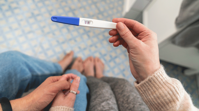
An ultrasound at 5 weeks isn’t the norm—there’s a good chance you may not even know you’re pregnant yet—but it does happen in special circumstances. Here’s why your provider might recommend a 5 week ultrasound.
A 5-week ultrasound isn’t a standard part of prenatal care and is “relatively uncommon,” says Catherine Caponero, DO, an ob-gyn at Cleveland Clinic. That’s because at 5 weeks pregnant, baby’s still too small to see much on a scan.
Although you’re only required to get one ultrasound during pregnancy, some practitioners perform first-trimester scans for all patients—often at around 8 weeks—to check for a heartbeat and estimate the due date.
Your doctor might recommend a 5-week ultrasound in “very specific circumstances,” says Caponero, including to follow up on abnormal bloodwork, severe pain, significant bleeding or if they’re concerned you may be at risk of an ectopic pregnancy or life-threatening miscarriage. “Some fertility doctors will perform ultrasound this early because they’re tracking Mom’s progress,” says Kimberly Khabeer, a sonographer at Temple University Hospital in Philadelphia.
I went in at 5 weeks, 5 days because of my earlier miscarriage. They saw a gestational and yolk sac but nothing else. It was cool to see because there was something there, but a little anxiety-provoking because it doesn't tell you much. We went back at 7 weeks and saw a strong heartbeat.
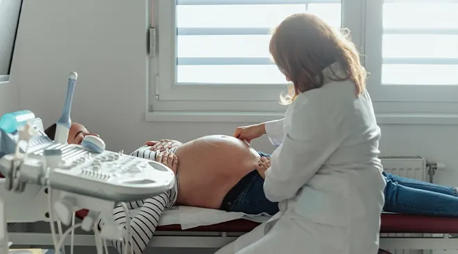
This early on, you can expect a transvaginal ultrasound: Your technician will place lubricant on an ultrasound wand, which they then insert into the vagina. (It might be a bit uncomfortable, but it shouldn’t be painful!) Ultrasounds typically take a few minutes, but it can take longer depending on what your practitioner needs to check. Good news: You don’t need to prepare in any way for a 5-week ultrasound.
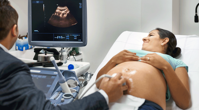
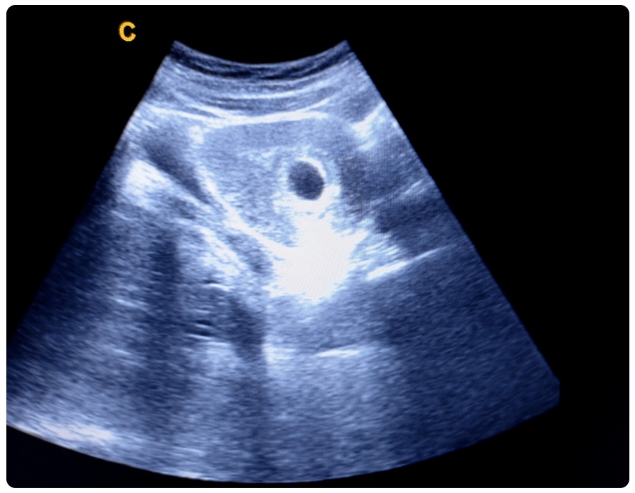
During a 5-week ultrasound, the sonographer will check for signs of pregnancy inside and outside of the uterus. You might see a very small gestational sac—a fluid-filled structure around the embryo—which resembles a dark-colored spot inside of the uterus. Inside the gestational sac, you might also spot a yolk sac, which nourishes the embryo until the placenta takes over around the end of your first trimester, says Caponero. “The yolk sac looks like a round or pear-shaped white circle with a black center on ultrasound,” she says.
Your practitioner usually won’t be able to see an embryo. They may also check the area around your ovaries and fallopian tubes for signs of an ectopic pregnancy, which is when a pregnancy implants outside of the uterus.
Don’t be alarmed if there’s no heartbeat—in fact, this is completely normal for a 5-week ultrasound, says Khabeer.
With my last pregnancy, I had an ultrasound around 5 weeks because of spotting and there wasn't a heartbeat. He was totally fine. I think it's pretty common to not be able to see or hear the heartbeat that early.
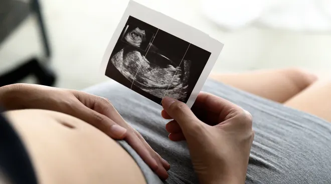
5-week ultrasound pictures
It’s true that you won’t be able to discern much on a 5-week ultrasound picture, but here’s what it might look like:
What if you see nothing at the 5-week ultrasound?
If you see nothing at your 5-week ultrasound, it’s not necessarily a problem. “Sometimes we can’t see anything,” says Khabeer. “It’s very possible to have a positive pregnancy test with no gestational sac visible on ultrasound at 5 weeks,” says Caponero. This may happen for several reasons, including that the pregnancy isn’t far enough along to be visible on scans. Other potential reasons include a chemical pregnancy (a pregnancy loss that happens before 5 weeks), a miscarriage or an ectopic pregnancy.
Because your doctor likely won’t see an embryo, they’ll usually schedule another ultrasound about 14 days later. “Just seeing a gestational sac isn’t enough. You need to see a fetal pole with cardiac activity to say it’s a viable pregnancy,” says Khabeer. This next ultrasound will also be able to confirm your estimated due date. “While it can be very hard to wait, it’s so important for expectant parents to have a clear answer instead of uncertain possibilities,” says Caponero.
At your next ultrasound, your practitioner should be able to detect an embryo with a heartbeat. If not, they may prescribe bloodwork to figure out the best time to repeat imaging, or to offer testing or treatment, Caponero says.
Frequently Asked Questions
Does an early ultrasound pose risks to Mom or baby?
Ultrasounds are very safe when performed by a trained and licensed practitioner. The main risk: Transvaginal ultrasounds can be a bit uncomfortable or awkward. Your doctor or technician will take steps to make you feel better, including using lubricant and letting you place the probe yourself, says Caponero.
Can you detect baby’s heartbeat at 5 weeks?
A heart is usually only visible at around 6 to 7 weeks of pregnancy. If there’s a heartbeat, it often means you’re actually further along. “In my experience, it's too early, and if you do [hear a heartbeat], the heart rate may be slow,” says Khabeer.
Can twins be detected at a 5-week ultrasound?
It may be possible to detect twins at a 5-week ultrasound. “You might see two sacs,” says Khabeer. But since you won’t be able to see the embryos, it's too early to confirm you’re actually carrying twins. It’s also possible you could be carrying twins that you can’t see just yet if they share the same amniotic sac, or because of where the pregnancy is located in the uterus and your anatomy.
Does an empty sac at 5 weeks mean a miscarriage?
No, an empty sac at 5 weeks doesn’t mean you’ve had a miscarriage. “It’s most likely too early to safely determine if it’ll be a successful pregnancy,” says Caponero. “This is why it’s so important to complete ultrasound or bloodwork follow-up as recommended by your provider to determine the best next steps.”
What are common pregnancy symptoms at 5 weeks?
Pregnancy symptoms vary a lot from person to person, but at 5 weeks pregnant, you may experience morning sickness, sore breasts, mood swings, a heightened sense of smell, a metallic mouth taste, light spotting and frequent urination, among other early pregnancy symptoms.
It’s unusual to get a 5-week ultrasound, because it’s simply too early to see much. You might get one if you have unusual symptoms like heavy bleeding and your practitioner wants to check for ectopic pregnancy or another serious condition. Basically, it’s a little extra peace of mind for your pregnancy journey—and that’s always welcome.
Please note: The Bump and the materials and information it contains are not intended to, and do not constitute, medical or other health advice or diagnosis and should not be used as such. You should always consult with a qualified physician or health professional about your specific circumstances.
Plus, more from The Bump:
Catherine Caponero, DO, is an ob-gyn at Cleveland Clinic. She earned her medical degree from Lake Erie College of Osteopathic Medicine in Erie, Pennsylvania.
Kimberly Khabeer is a sonographer at Temple University Hospital in Philadelphia.
American College of Obstetricians and Gynecologists, Ultrasound Exams, January 2024
Cleveland Clinic, Transvaginal Ultrasound, May 2022
Cleveland Clinic, Ultrasound in Pregnancy, September 2022
Cleveland Clinic, Yolk Sac, February 2022
Medline Plus, National Library of Medicine, Ultrasound Pregnancy, March 2024
National Health Service UK, Week 5
Penn Medicine, Level II Ultrasound
StatPearls, National Library of Medicine, Gestational Sac Evaluation, July 2023
StatPearls, National Library of Medicine, Sonography 1st Trimester Assessment, Protocols, and Interpretation, July 2023
Real-parent perspectives
- Bookhousegirl, The Bump community member
- DRB7586, The Bump community member
Learn how we ensure the accuracy of our content through our editorial and medical review process
Navigate forward to interact with the calendar and select a date. Press the question mark key to get the keyboard shortcuts for changing dates.