


What to Expect at the 14-Week Ultrasound Appointment

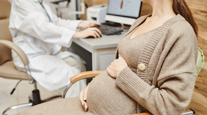
Your 40 or so weeks of pregnancy will have a lot of prenatal appointments in store for you—more if you’re high-risk. Sprinkled throughout these appointments, you’re likely to have a few pregnancy ultrasounds to see what your little one’s up to and how they’re developing. If you’re approaching the early weeks of your second trimester, you may be wondering whether or not to expect an ultrasound at 14 weeks. The short answer: It depends. Ahead, read why your provider might order a 14-week ultrasound, and what you can expect to see on screen.
Once you’ve reached 14 weeks of pregnancy, you’ve likely already had at least one ultrasound, if not two. “For an uncomplicated pregnancy, there’s usually an ultrasound in the first trimester,” says Julia Timofeev, MD, a board-certified ob-gyn and maternal-fetal medicine specialist in Washington, DC. She says this ultrasound usually happens around 11 to 13 weeks, and it’s used to assess fetal growth and confirm baby’s due date.
Pregnant women typically also have the option to have a nuchal translucency scan between weeks 11 and 13, which includes an ultrasound. While this scan is typically performed within the 11- to 13-week window, some providers will also perform it at 14 weeks, which would mean getting an ultrasound at this stage.
Another reason someone may get an ultrasound at their 14-week appointment is if they weren’t able to be seen during their first trimester and they’re behind on their prenatal care. Otherwise, getting a 14-week ultrasound isn’t usually part of the standard schedule for an uncomplicated pregnancy.
According to Katherine R. Goetzinger, MD, a board-certified ob-gyn and maternal-fetal medicine specialist in Baltimore, Maryland, after the first trimester and/or nuchal translucency scan ultrasounds, the second ultrasound is most often the anatomy scan, performed between 18 to 20 weeks of gestation. The American College of Obstetricians and Gynecologists (ACOG) recommends at least one ultrasound during pregnancy, typically the anatomy scan.
While a 14-week ultrasound isn’t standard, there are certain reasons your provider may recommend it. “An expectant patient’s ultrasound schedule is determined based on their personal medical history and past obstetrical history, as well as the expectant couple’s family history, including any risks for congenital defects or genetic conditions,” says Alice Cootauco, MD, a board-certified ob-gyn and maternal-fetal medicine specialist in Baltimore, Maryland.
Some reasons Cootauco says your provider might recommend an ultrasound at 14 weeks may include:
- To screen for congenital anomalies in a patient with increased risk
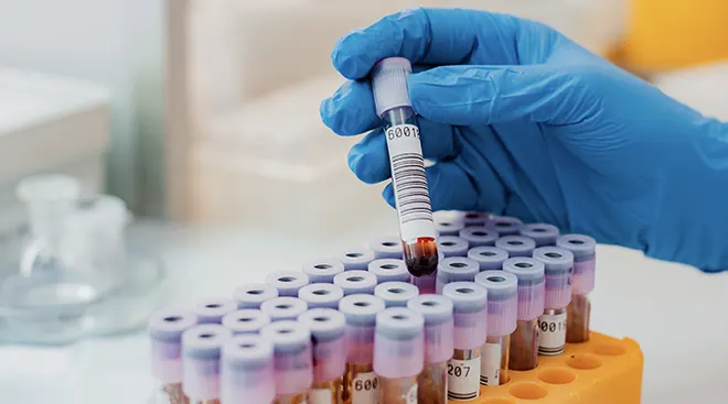
- To screen for chromosomal defects, potentially as part of an aneuploidy screening (which may include a nuchal translucency scan)
- To detect chromosomal or genetic abnormalities with chorionic villus sampling (CVS) (typically due to abnormal aneuploidy screening or if the fetus is at risk for an inherited genetic condition)
- To better pinpoint baby’s gestational age if the date of your last menstrual period is unknown, or if there’s a discrepancy between your uterine size and baby’s due date
- To confirm the presence of twins or multiples, especially if your uterine size doesn’t match with the suspected due date
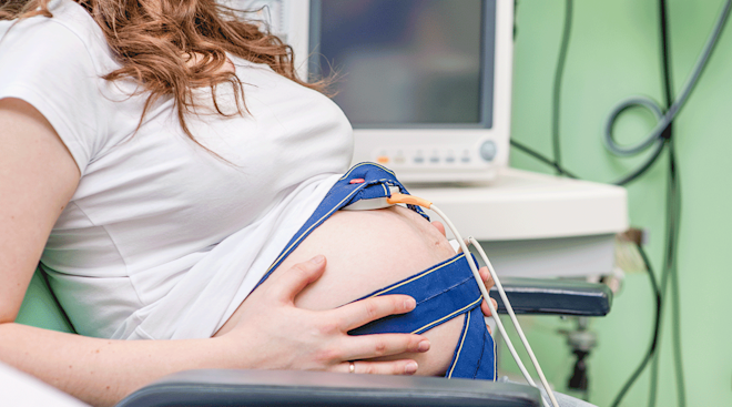
Timofeev notes that an ultrasound at 14 weeks may be performed as a follow-up to a first-trimester scan with concerning findings, such as “to reassess fetal heart rate in cases of early pregnancy bleeding, or to follow up on interim fetal growth.”
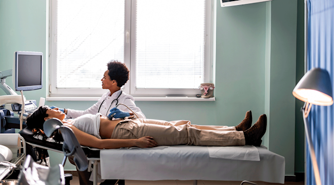
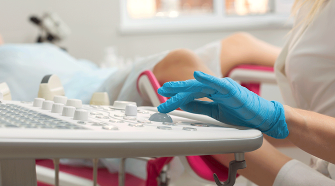
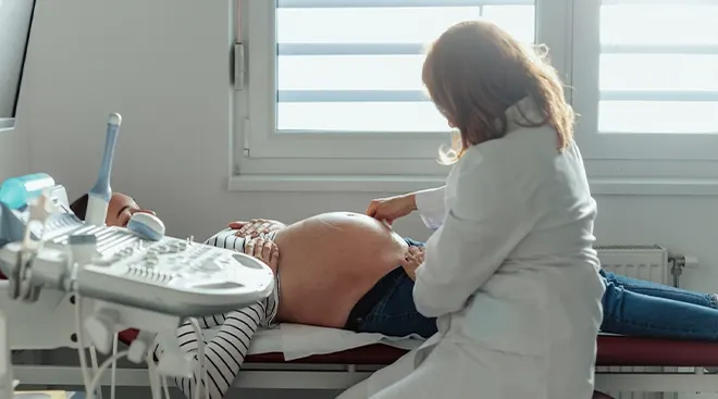
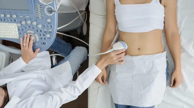
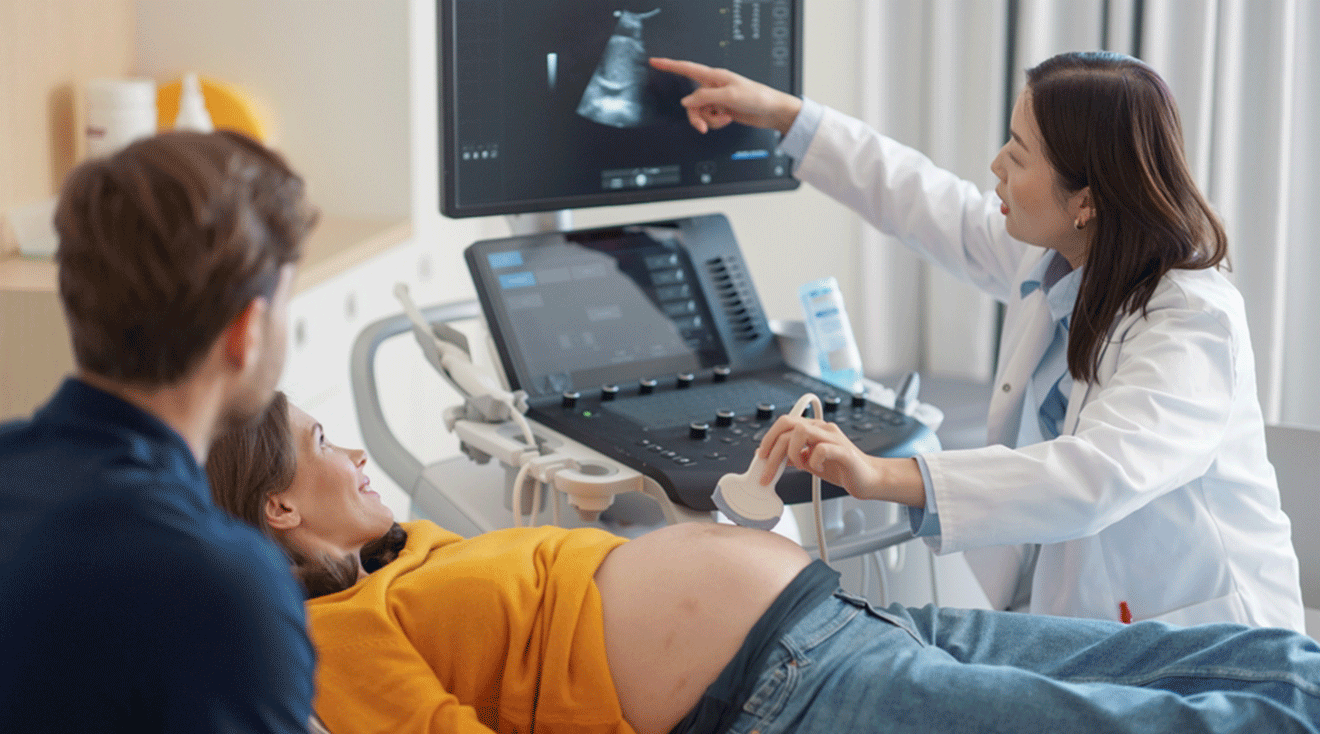
If you’re having an ultrasound at 14 weeks, it will most likely be transabdominal, meaning your provider will put some gel on your belly (which may feel cold) and then spread it around with the handheld transducer to get a look at baby. In some cases, however, you may end up having a transvaginal ultrasound, where the transducer is wand-shaped and inserted vaginally. “The approach depends on the specific reason the ultrasound is being performed, as in some cases it may be difficult to visualize pregnancy clearly on transabdominal ultrasound,” explains Timofeev.
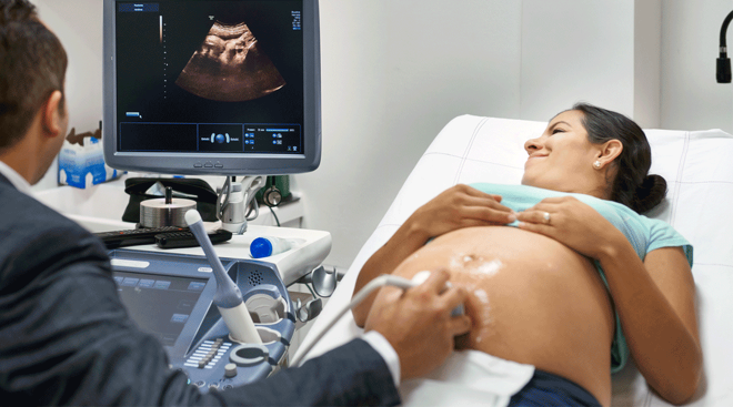
Either way, your provider will be able to get a look at baby, and you’ll get to watch as they move the transducer around to get different views. You might see your little one moving around during the scan, and you’ll be able to see a profile that looks like a baby (unlike during an early first-trimester scan, when they still looked like a peanut).
During the scan, your provider will freeze the video periodically to take measurements of baby and your uterus. Most of the time, your provider will also freeze the video when they have a really good image of baby so that they can print the image for you to take home.
While it’s unlikely to get a 100 percent answer on baby’s sex right now, it’s possible. My 14-week ultrasound showed that my little one was definitely a boy! Of course, we were lucky enough to see him at just the right angle to clearly see his little boy parts. If they don't see much, they'll tell you they aren't certain and that you may have to wait.
By the time you’ve reached 14 weeks pregnant, baby can wiggle their fingers and toes, move their head from side to side, suck their thumb and make facial expressions. At a 14-week ultrasound, Timofeev says providers can also see whether you’re having twins or multiples, though this will likely have been detected earlier. Baby’s sex may also be visible, although she notes that any ultrasound-based sex determination isn’t as accurate as it will be during your anatomy scan later on.
Additionally, Goetzinger says most of baby’s organ systems are formed by 14 weeks, and providers can usually “identify major malformations, such as heart defects or brain abnormalities.” Cootauco further explains, “In general, 50 to 70 percent of major congenital anomalies [and] approximately 90 percent of lethal anomalies can be detected before 14 weeks.”
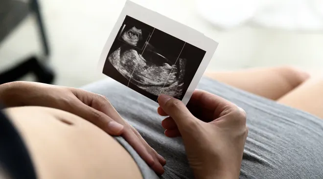
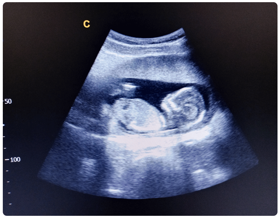
14-week ultrasound pictures
If you find yourself getting an ultrasound at 14 weeks, chances are high that you’ll get to go home with some pictures of your little one. Even better, baby will have an identifiable profile, so it’ll look like a real infant, not just an oddly shaped dark spot. Here’s an example of an ultrasound photo at 14 weeks.
Frequently Asked Questions
How big is baby at this stage?
At 14 weeks, baby’s about as big as a peach. They likely measure around 3.5 inches and weigh about 1.5 ounces. In fact, they’re big enough that your bump might start showing soon if it hasn’t already.
How accurate is sex determination at a 14-week ultrasound?
Again, while baby’s genitalia may be visible at 14 weeks, it’s usually not clear enough for providers to be 100 percent sure of their sex. Your anatomy scan at 18 to 20 weeks will determine baby’s sex a lot more accurately. If you'd like to find out earlier, noninvasive prenatal testing (NIPT) can tell you as early as 10 weeks of pregnancy.
Can twins go undetected at 14 weeks?
Usually, you can see twins on an ultrasound by 12 weeks. By the time you reach 14 weeks, if you have an ultrasound, Goetzinger says, “You can certainly identify if there are multiples.”
Can I still get the nuchal translucency test at a 14-week ultrasound?
According to our experts, the ideal time to get a nuchal translucency test is between weeks 11 and 13. “There are strict criteria in place so that the nuchal translucency measurements are highly precise and informative,” says Timofeev. Depending on baby’s size, you may still be able to get the test at 14 weeks. But “if the measurement of the fetus exceeds a certain threshold,” the test may not be accurate, Goetzinger notes.
In most low-risk, uncomplicated pregnancies, a 14-week isn’t standard, so if you’re all caught up on appointments at this point, you can expect your next scan to come between 18 and 20 weeks. However, every pregnancy is different, so if your doctor recommends a scan at 14 weeks, it’s best to follow their guidance.
Please note: The Bump and the materials and information it contains are not intended to, and do not constitute, medical or other health advice or diagnosis and should not be used as such. You should always consult with a qualified physician or health professional about your specific circumstances.
Plus, more from The Bump:
Alice Cootauco, MD, is a maternal-fetal medicine specialist with expertise in fetal diagnosis and management of maternal and pregnancy complications, and the director of the Perinatal Care Center at the University of Maryland St. Joseph Medical Center. She earned her medical degree from Emory University.
Katherine R. Goetzinger, MD, MSCI, is a maternal and fetal medicine physician, associate professor, program director of the maternal-fetal medicine fellowship program and director of perinatal outreach at the University of Maryland School of Medicine. She earned her medical degree from the University of Maryland School of Medicine.
Julia Timofeev, MD, FACOG, is a board-certified ob-gyn and maternal-fetal medicine specialist, and the medical director at MFM & Genetics of Advantia in Washington, DC. She earned her medical degree from the University of Florida College of Medicine.
American Academy of Family Physicians, Testing for Fetal Aneuploidy, April 2020 American College of Obstetricians and Gynecologists, Ultrasound Exams, January 2024
National Health Service UK, Pregnant with Twins, October 2022
Real-parent perspectives
Learn how we ensure the accuracy of our content through our editorial and medical review process.
Navigate forward to interact with the calendar and select a date. Press the question mark key to get the keyboard shortcuts for changing dates.